What is a secondary antibody?
Secondary antibodies bind to antibodies or antibody domains from other species. They are used for indirect detection of the target antigen by binding to the primary antibody. Where primary antibodies bind specifically to an epitope of the antigen, secondary antibodies bind to the conserved regions of an antibody. These conserved regions are constant within a particular class of antibodies such a mouse IgG. Meaning that a secondary antibody can be used with any primary antibody of the same host species. Making secondary antibodies more versatile and cost reducing than an individually labeled primary antibody.
How is a secondary antibody produced?
Secondary antibodies are produced by immunizing a host animal with target antibodies from another species. For example, to produce anti-mouse secondary antibodies, a host animal will be injected with mouse IgG. Immunizing a rabbit with mouse-IgG, the rabbit will generate rabbit anti-mouse IgG secondary antibodies. Common used animals for production of secondary antibody are goat, donkey and rabbit but other animals can also be used. The selected secondary antibody should match the species in which the primary antibody was raised and the isotope of the primary antibody that is targeted. A rabbit anti-mouse IgG secondary antibody can be used for binding a mouse IgG primary antibody. Because of the high degree of conservation between immunoglobulins it is necessary to purify the secondary antibodies with affinity purification to achieve minimal cross-reaction with other immunoglobulins.
Secondary antibody formats
Whole IgG secondary antibodies
These secondary antibodies are intact molecules, purified from antisera with affinity chromatography. They have an Fc portion and 2 antigen binding Fab portions joined together by non-covalent interaction. The whole IgG form of antibodies is suitable for the majority of immunodetection procedures.
F(ab)2 fragment secondary antibodies
The F(ab)2 antibody fragments are created by digestion of the whole IgG antibodies. With pepsin the Fc fragment of the antibody is removed, while leaving the hinge region intact. The F(ab)2 fragments still have 2 antigen binding sites. These fragments are used in specific applications. When working with live cells, the use of these F(ab)2 fragments can prevent binding of secondary antibodies to the Fc receptors on the cell surface.
Movalent Fab Fragment secondary antibodies
Fab fragment antibodies are also created by digestion of the whole IgG antibody. Digestion with papain removes the Fc fragment, including the hinge region. Creating fab fragments with a single binding site. The Fab fragments can be used to block endogenous immunoglobulins on the cell surface or block the surface of immunoglobulins for doble labeling primary antibodies from the same species.
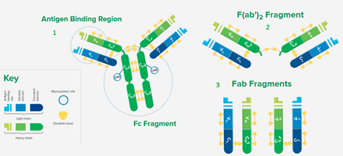
Figure 1: 3 antibody formats; 1:whole IgG secondary antibody, 2: F(ab)2 Fragment antibody, 3: Fab Fragment
Advantage of secondary antibodies; direct vs indirect detection
Primary antibodies have limited binding capacity for antigens. Multiple conjugated secondary antibodies can bind to a single primary antibody. Causing signal amplification and therefore increasing the sensitivity of the application. To increase the signal amplification even more, a biotinylated secondary antibody can be used in combination with conjugated streptavidin.
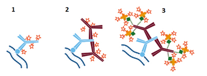
Figure 2: direct vs indirect detection; 1: direct detection with labeled primary antibody, 2: indirect detection with labeled secondary antibody, 3: indirect detection with biotinylated secondary antibody and conjugated streptavidin.
Secondary antibodies can be used unconjugated as capture antibody, but they can also be conjugated with a great variety of labels such as fluorophores, enzymes or proteins to amplify the signal of primary antibodies in a wide range of applications, including ELISA, Western blotting, Immunostaining and Flow Cytometry.
Our supplier Jackson ImmunoResearch is specialized in secondary antibodies and conjugates. They offer more than 3.000 secondary antibodies for detecting primary antibodies in all applications, including ELISA, Eltectron microscopy, Western Blot, Flow Cytometry and Confocal microscopy (Immunohistochemistry, Immunocytochemistry and Immunofluorescence).