How to choose the right microscope setup for your experiment

All scientists need to obtain convincing images to support the biological phenomenon they are studying. The main question is how to choose the best microscope setup to answer the experimental question in the most efficient manner.

FLUORESCENCE MICROSCOPY: WIDEFIELD VERSUS CONFOCAL MICROSCOPY

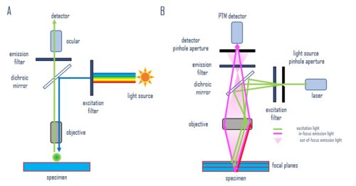
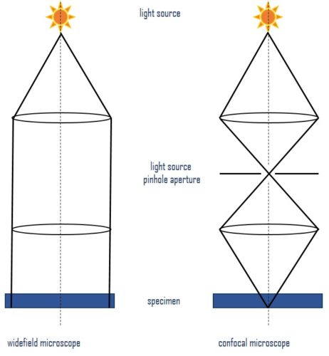
Fluorescence microscopy is an imaging technique that uses fluorescent probes, dyes, or proteins (fluorophores) for sample labeling to generate an image. Which microscope you choose will depend on the individual experiment. Widefield microscopes are more robust and provide a general picture quickly. While confocal microscopes should be used for thicker samples, or higher-resolution pictures (Figure 1).
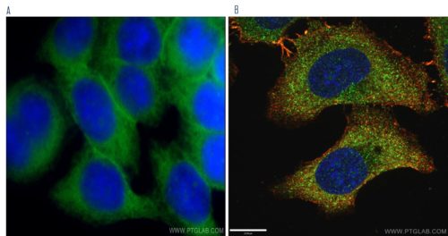
SLC2A1, GLUT1 antibody

Figure 1.
A) Immunofluorescent analysis of HeLa cells using SLC2A1, GLUT1 antibody (21829- 1-AP) at a dilution of 1:50 and Alexa Fluor 488-conjugated AffiniPure Goat Anti-Rabbit IgG(H+L) by widefield microscope.
B) Immunofluorescent analysis of (-20°C Ethanol) HepG2 cells fixed using LAMP1 antibody (55273-1-AP) at a dilution of 1:50 and Alexa Fluor 488-conjugated AffiniPure Goat Anti-Rabbit IgG(H+L) by confocal microscope. DAPI was used as the nuclear counterstain.

THE MAIN DIFFERENCE: CONFOCAL - WIDEFIELD MICROSCOPY

A widefield microscope (Figure 2A) has a specially selected excitation light, which illuminates the specimen through the objective lens. The fluorescence emitted by the labeled specimen is focused on the detector by the same objective that is used for the excitation light. The dichroic mirror acts as a wavelength-specific filter, transmitting fluorescence through to the eyepiece or detector but reflecting any remaining excitation light toward the source.

A confocal microscope (Figure 2B) uses a light band emitted by a laser. A specific light wavelength passes through a pinhole and is reflected by the dichroic mirror toward the specimen, which is scanned on a selected focal plane. The excited fluorophore emits secondary fluorescence that passes through the dichroic mirror and is focused as a confocal point at the photomultiplier (PTM) detector pinhole.

Widefield and confocal microscope


Figure 2. Image generation in widefield epifluorescence (A) and confocal (B) microscopes.

The main difference between confocal and widefield microscopy is the use of a pinhole in a confocal microscope. It allows only light from the plane of focus to reach the detector. This reduces the acquisition of out-of-focus light, improving image quality (Figure 3).

Light paths in microscopy

Figure 3. Light paths in widefield and confocal microscopy – visualization of the pinhole principle.

USE A MICROSCOPE RELEVANT TO ANSWERING YOUR QUESTION

For most cell biology applications, a widefield fluorescence microscope is suitable and provides a good trade-off between image quality, speed of acquisition, and cost. It is most applicable for:

-              imaging tissue culture cells or other thin specimens;
-              initial protocol screens and verification of the staining quality before using confocal microscopy or other technologies;
-              live cell imaging where speed of acquisition is an advantage over scanning using confocal-based approaches.

A confocal microscope would be better for:

-              subcellular localization studies,
-              protein–protein interactions and colocalizations,
-              3D imaging of thick tissues,
-              polarized cells,
-              larger specimen surfaces (stitching between fields),
-              multi-fluorescence imaging,
-              time-lapse imaging,
-              FLIM (fluorescence-lifetime imaging) and FRAP (fluorescence recovery after photobleaching) measurements.

The crucial question before starting is “what do I want to see?” The answer should determine the experimental setup, sample preparation, and imaging platform used.