Organoid Culture

Combining the principles of developmental biology with stem cell culture techniques to grow biological tissues in a dish

Researchers have devised methods to generate stable, physiologically relevant miniature organ models that simulate brain, liver, thymus, thyroid, lung, pancreas, and heart tissue. In addition to providing a detailed view of how organs form and grow, these models—called organoids—are used for drug discovery and toxicology research, infectious disease modeling, and tissue engineering/gene editing for regenerative medicine, providing new pursuits for developing personalized therapeutics from individualized tissues.

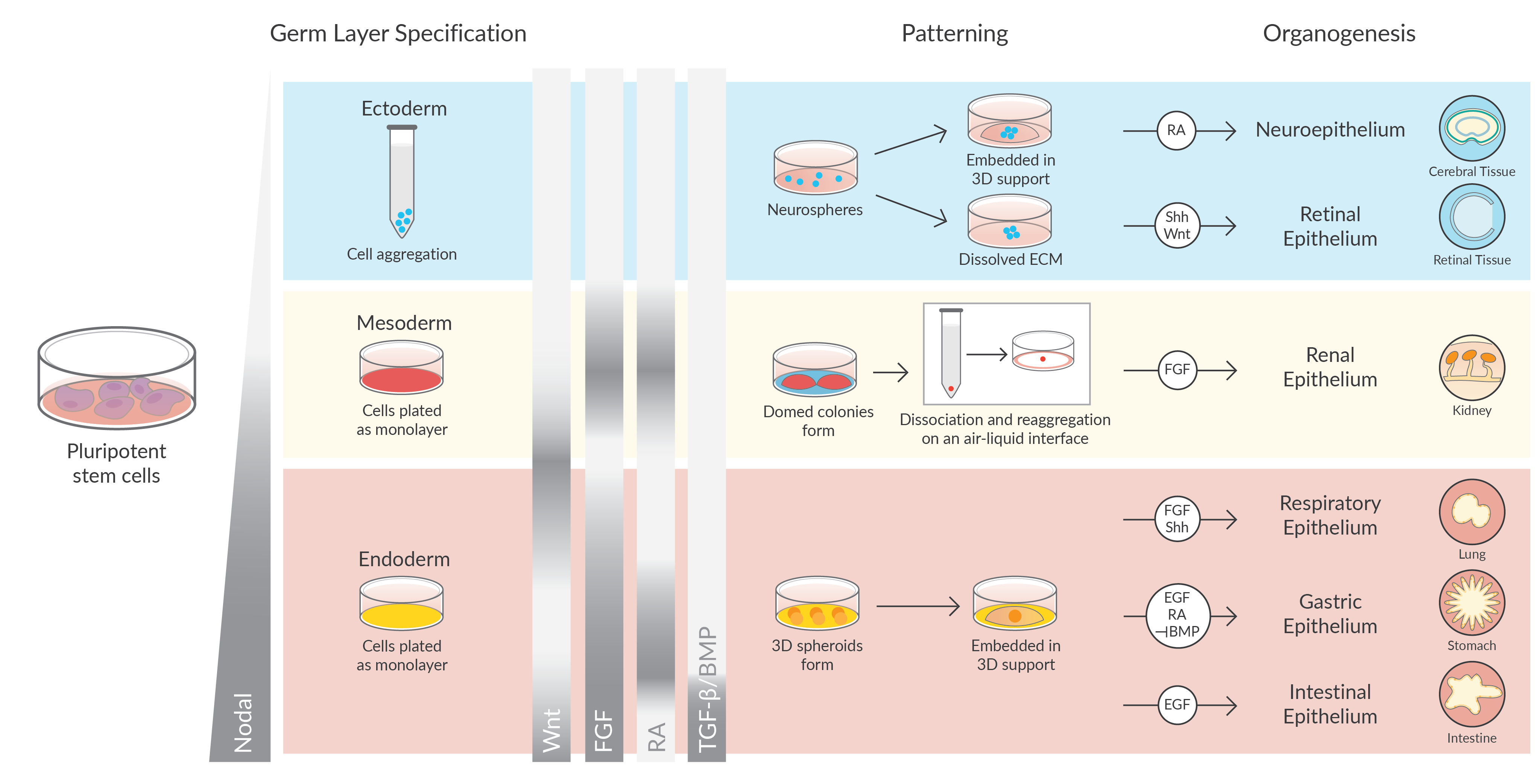
Organoids are tiny, three-dimensional cultures that are derived from tissue or pluripotent stem cells. Using the knowledge gained from maintaining stem cell populations, the cells are cultured in an environment that allows them to follow their own genetic instructions to self-organize into tissue with some organ-like functionality that contains a self-renewing stem cell population. Self-assembly and differentiation are the result of instructive signaling cues given to the cells by the extracellular matrix, the culture medium, and, by the cell types present in the organoids themselves—once the structure assembles.

Culturing Organoids

Different tissues require their own specific culture methods, but generally pluripotent stem cells or tissue-specific progenitor cells are embedded in Matrigel®, or similar extracellular matrix, and grown in the presence of cell culture media containing specific growth factors that mimic the in vivo signals required for maintenance of the stem cell population. Under these conditions, embedded cells proliferate and self-organize into three-dimensional organoid structures that can be passaged and maintained indefinitely. Exposing these cells to specific combinations and concentrations of growth factors and other signaling molecules triggers differentiation and morphogenesis (Figure 1).

Organoid Culture

Figure 1. Germ layer specification, patterning, and organoid development are governed by shared developmental cues. Image adapted from Development 144(6), 958-962 (2017).

Wnt, FGF, retinoic acid (RA), and TGF-β/BMP are the main patterning molecules that govern germ layer formation, patterning, and organogenesis. During gastrulation, epiblast cells migrate through the primitive streak, segregating the mesoderm and endoderm from the ectoderm. Nodal, a member of the TGF-β superfamily is required for mesoderm and endoderm formation where a short exposure to nodal leads to a mesendodermal fate and longer exposure (higher levels) of nodal influence the formation of definitive endoderm. Nodal and Wnt signaling are essential for gastrulation to occur in the posterior epiblast, yet repression of these pathways in the anterior epiblast is essential for neuroectoderm formation.

Neural and Retinal Patterning

A defining feature of directing the differentiation of pluripotent stem cells into neural tissues is the absence of inductive signals. Using this concept, neural cells have been differentiated in culture under the influence of small molecule Wnt inhibitors. However, once neural identity is established the neuroepithelium requires patterning factors to form organ-like structures. For instance, defined cerebral regional domains arise in the presence of RA. On the other hand, retinal epithelial tissues differentiate in the presence of sonic hedgehog (Shh) and Wnt.

Renal Patterning

In mesoderm, the timing of the migration of presomitic mesoderm through the primitive streak can be modeled by the sequential activation of Wnt then FGF signaling. This results in anterior-patterned and posterior-patterned intermediate mesoderm that can generate ureteric epithelium and metanephric mesenchyme, respectively. Ureteric epithelial identity is also promoted by exposure to RA, whereas metanephric mesenchyme arises in the absence of RA signaling. Prolonged exposure to Wnt and FGF further promotes growth of kidney organoids.

Fore/Mid/Hindgut Patterning

Spatial and temporal gradients of Wnt, FGF, RA, and TGF-β/BMP control endoderm patterning along the anterior-posterior axis. In addition to these four signals, posterior endoderm identity is determined by the transcription factor Cdx2. Activation of Wnt and FGF signaling promotes the expression of CDX2, resulting in a commitment to mid/hindgut fate. In conjunction with Wnt and FGF activation, anterior endoderm patterning requires inhibition of BMP signaling to repress Cdx2 (posterior programming) and instead promote the formation of Sox2-expressing foregut endoderm. The posterior portion of the foregut is patterned by exposure to RA, with continued activation of Wnt signaling enabling the production of gastric organoids. Anterior foregut endoderm, patterned by the inhibition of TGF-β/BMP, gives rise to respiratory epithelium. Once patterned, endodermal organoids are supported by tissue-specific factors. Epidermal growth factor (EGF) is required to maintain gastric and intestinal organoids, while FGF and Shh are required to promote lung epithelial development.

Resources for Organoid and 3D Cell Culture

Media formulations necessary for deriving and sustaining organoids from epithelial tissues include small molecules that potentiate Wnt pathway activity, BMP signaling antagonists, and several additional factors. See the list below for a compilation of matrix and media components available from Cayman.

Product Name Matrix Component Media Component Use in Culture
A 83-01 TGF-β inhibitor added for long-term organoid growth
L-Alanyl-L-Glutamine  √ Stable L-glutamine dipeptide that enables trophic expansion
CHIR99021  √ Wnt/β-catenin pathway activator required for proliferation
DAPT  √  √ Indirect Notch pathway inhibitor; important for intestinal organoid differentiation
Forskolin  √  √ Forskolin-induced swelling of intestinal organoids
Gastrin I (human)  √ Establish stomach, intestine, colon, and liver organoid cultures
IWP-2  √ Wnt/β-catenin pathway inhibitor promotes heart organoid differentiation
IWP-4  √ Wnt/β-catenin pathway inhibitor promotes heart organoid differentiation
IWR-1-endo  √ Wnt/β-catenin pathway inhibitor promotes cerebral organoid differentiation
LDN-193189 BMP type I receptor inhibitor important for proliferation and brain organoid differentiation
LDN-193189 (hydrochloride) BMP type I receptor inhibitor important for proliferation and brain organoid differentiation
Nicotinamide Culture supplement for long-term organoid growth
PD 0325901 MEK/ERK inhibitor maintains cell proliferation
Prostaglandin E2 Promotes sustained growth and survival of epithelial spheroids as an alternative to Wnt agonists
all-trans Retinoic Acid Potent, time-dependent caudalizing factor of the neuroectoderm and neuronal and gastric epithelium differentiation-inducing molecule
SB 202190 p38 MAPK inhibitor added for long-term growth; supports gastric organoid culture
SB 202190 (hydrochloride) p38 MAPK inhibitor added for long-term growth; supports gastric organoid culture
SB-431542 (hydrate) TGF-β/Activin/NODAL pathway inhibitor that promotes brain, liver, and blood vessel organoid differentiation
Y-27632 (hydrochloride) ROCK inhibitor used for long-term maintenance and cryopreservation of organoids