Markers of neuronal precursor cells (NPCs) / neural stem cells (NSCs)

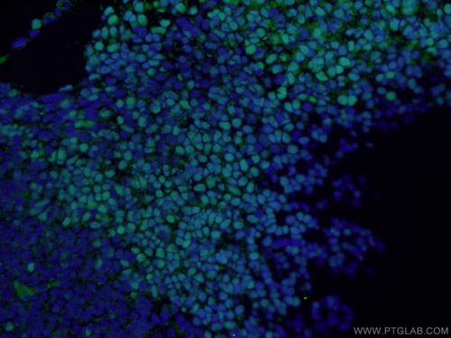
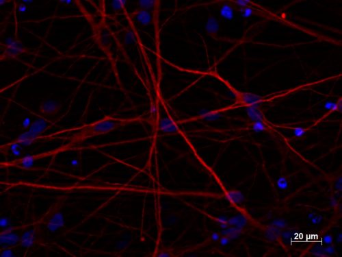
What are neural stem cells (NSCs), neural progenitors, and neural precursors (NPCs)?

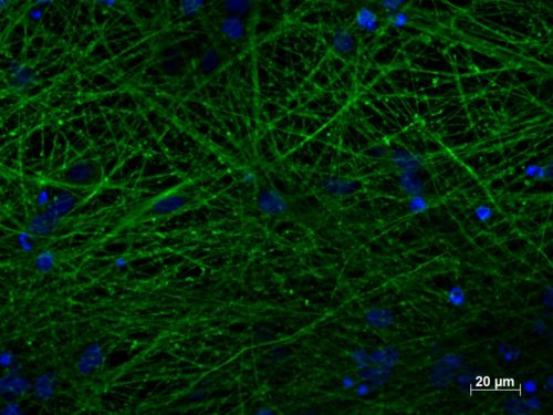
Neural stem cells (NSCs) are undifferentiated neural cells with the capacity for long-term self-renewal and for differentiation into all types of neuronal and glial cells. They play a crucial role in shaping the nervous system during development but are also important in adulthood.

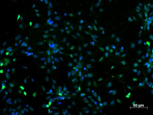
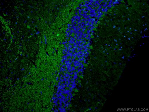
A subset of NSCs is present in the developed nervous system and acts as a reservoir of cells for cell replacement and nervous tissue regeneration (PMID: 20110496). Symmetric division of NSCs maintains the population of undifferentiated NSCs, while asymmetric division gives rise to neural progenitors. Neural progenitors are precursor cells able to further differentiate into various neuronal and glial cells. However, unlike NSCs, they have limited life time and are not able to self-renew. NSCs and neural progenitors are often regarded as neural precursors (NPCs). Adult NPCs are particularly enriched in the subventricular zone, in the dentate gyrus, and potentially also in the hypothalamus.

NPC Markers

NPCs are distinct from differentiated cells of the nervous system at the molecular level. They differ in the expression of transcription factors and plasma membrane proteins. They can be used as NPC markers for immunohistochemistry and flow cytometry analysis. However, NPCs are quite heterogeneous and there are distinct subpopulations of NPCs within the nervous system (PMID: 24362763).

Hippocampal NPCs express glial fibrillary acid protein (GFAP), sex determining region Y-box 2 (SOX2; Figure 1), brain lipid-binding protein (BLBP), and nestin (PMID: 21549330). Proliferative NPCs are characterized by the expression of SOX2 and paired box 6 (PAX6; Figure 2). Doublecortin (DCX; Figure 3) or PSA-NCAM are markers frequently used for intermediate progenitor cells and early immature neurons (PMID: 29625071). Mature neurons are positive for neuronal nuclei (NeuN), tubulin beta 3 class III (TUBB3; Figure 4), and microtubule-associated protein 2 (MAP2; Figure 5) (PMID: 26931327).

SOX2 Antibody

Figure 1. Immunofluorescent analysis of (4% PFA) fixed mouse embryo tissue using 20118-1-AP (SOX2 antibody) at dilution of 1:50 and Alexa Fluor 488-conjugated AffiniPure Goat Anti-Rabbit IgG(H+L)

PAX6 Antibody

Figure 2. Immunofluorescent staining of PAX6 (12323-1-AP, 1:250 dilution) with 4% PFA fixed control human-induced pluripotent stem cell (hiPSC)-derived NPCs. (Red: PAX6; Blue: DAPI). Provided by BioTalentum Ltd., Hungary.

DCX Antibody

Figure 3. Immunofluorescent analysis of (4% PFA) fixed mouse brain tissue using 13925-1-AP (DCX antibody) at dilution of 1:50 and Alexa Fluor 488-conjugated AffiniPure Goat Anti-Rabbit IgG(H+L)

TUBB3 Antibody

Figure 4. Immunofluorescent staining of TUBB3 (66375-1-lg, 1:250) with 4% PFA fixed control hiPSC-derived neuronal cultures (35 days old). (Green: TUBB3; Blue: DAPI). Provided by BioTalentum Ltd., Hungary.

MAP2B Antibody

Figure 5. Immunofluorescent staining of MAP2 (17490-1-AP, 1:250 dilution) with 4% PFA fixed control hiPSC-derived neuronal cultures (35 days old). (Red MAP2; Blue: DAPI). Provided by BioTalentum Ltd., Hungary.

In vitro NPC cultures

NPC markers are also useful in establishing and monitoring in vitro NPC culture differentiation. NPCs can be differentiated from embryonic stem cells (ESCs) but also from induced pluripotent stem cells (iPSCs) (PMID: 11731782). NPCs can additionally be obtained by direct reprogramming of somatic cells from different cell lineage (PMID: 22445518). This approach is particularly useful for obtaining patient-specific NPCs used as established disease models (PMID: 28105055). Alternatively, NPCs can be isolated from fresh nervous tissues using cell sorting with NPC markers (PMID: 11121071). NPCs are typically grown in vitro in the form of neurospheres or as adherent monolayers (PMID: 26798357).